American foulbrood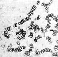
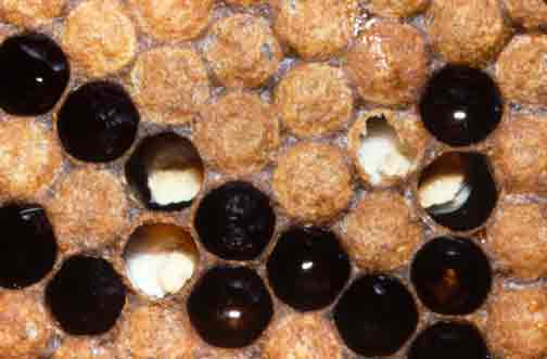
Clinical manifestation: changes on hatches (dark gray with holes), on larvae (pure white, they die and turn into liquid chocolate-like dark mass), and in brood itself (fewer larvae). Dead bees can also be found in hive
Treatment: disease isn't treated. Methods of eradication: burning, mechanical cleaning, sanitary washing and disinfection. Note: in most countries the state is refunding damages to beekeepers if the disease is not older than 2 months (black liquid chocolate like mass in cells). The disease is older than 2 months if the cells are dry and empty. Consult a local veterinarian for details. Diagnosis: material for examination: carved out honeycomb (4x4in-10x10cm, 4x8in-10x20cm or entire frame). The sample is to be packed in clean paper, properly marked and then sent to a laboratory with accompanying act and anamnestic data. |
New additions
Now you can have all the bee treatment details with you,with bee diseases app from google play. |
Microscopic exam: With toothpick (or matches) wood dark matter is to be extracted from cells and placed on a microscopic plate. The material is then dried, fixated and colored. Vegetative forms coloring: gram, gimza, methylen blue (can be detected in the first days of infection). Spores are colored with carbol fuchsin (they are spotted in pairs or as singles - resemble top part of the needle, transparent bodies, red layer). Nutritive mediums: standard or enriched. Note: pestis has bactericide properties, so other microorganisms wont grow on the medium.
Pathoanatomical changes: in clinical picture.
Epizootology and etiology: Caused by 'Paenibacillus larvae', very contagious, hard to eradicate. Vegetative forms look like sticks, can create spores, extremely durable (can survive 20-30 years).
Pathoanatomical changes: in clinical picture.
Epizootology and etiology: Caused by 'Paenibacillus larvae', very contagious, hard to eradicate. Vegetative forms look like sticks, can create spores, extremely durable (can survive 20-30 years).
European foulbrood
Clinical manifestation: brood weakens, its hatches are dark gray and damaged. There are dead larvae in the cells.
Treatment:sugar syrup, antibiotics and dealing with unspecific factors - unhygienic beekeeper or community without queen-bee.
Diagnosis: samples- part of the honeycomb (4x4in-10x10cm, 4x8in-10x20cm). It must be properly packed and marked. Samples are then derived from larvae and mixed with saline solution and colored by next methods: gram, gimza, methylen blue, gentian violet. Positive findings: g+ and g- sticks, coccus, spores, spores of 'bacillus alvei' (ellipsoidal shape). Nutritive medium- usual mediums.
Pathoanatomical changes: in clinical picture.
Epizootology and etiology: mixed infection of a complicated etiology - bacillus alvei, streptococcus faecalis, streptococcus pluton. Other factors: unhygienic beekeeper and community without a queen-bee. This disease has an explosive character.
Epizootology and etiology: mixed infection of a complicated etiology - bacillus alvei, streptococcus faecalis, streptococcus pluton. Other factors: unhygienic beekeeper and community without a queen-bee. This disease has an explosive character.
Stone brood
Clinical manifestation: larvae die and their bodies become solid like a rock. If the frame is shaken, crumbling stone-like noises can be heard. Brood color turns green like it's been covered with moss.
Treatment: this disease is a zoonosis (it can affect humans - complicated pneumonia (death if not treated)) and thus is not treated. If this disease is discovered a veterinary inspection should be contacted immediately in order for hives to be safely destroyed (most often by burning). Bee keepers should not be worried though, most states are refunding damage caused by this disease. When the bees are destroyed, hives can be cleaned mechanically and with disinfectants. After that they are washed with hot water and are left to dry. Honey from from supper can be used in the confectionery industry after boiling (and mixing with water). During all these times protective equipment should be used (mask, suit and gloves). Note - regardless of what I just wrote here (these are the opinions of the scientific community) I would proceed to burn it all nonetheless since this disease is a zoonosis. Always bear in mind the fact that if any mistake is made you too can get infected.
Diagnosis: material for examination includes: larvae and part of the honeycomb (4x4in-10x10cm, 4x8in-10x20cm). It must be properly packed and marked, microscopic or via the nutritive medium. For microscopic examination parts of bee (or larvae) carcasses are taken and sunk into 50% solution of glycerin or NaCl. After that sample is observed natively (directly under the microscope). Condiospores with sterigmas can be spotted. For examination with the nutritive medium special mediums are used (Sabouraud agar). Again when working with A. flavus care should be taken since it causes complicated pneumonia in humans.
Pathoanatomical changes: in clinical picture.
Epizootology and etiology: caused by fungus 'asperegilus flavus link', 'asperegilus niger' or 'asperegilus fumingatus'. Unspecific factors are also important for disease development. This disease causes Asperegilosis in bees. Favorable factors for disease are high moisture in hive and weak societies. Spores are found anywhere in the hive and on the bees (they can thus spread disease to another hive). Bees are infected by ingesting spores. Fungus Asperegilus flavus Link - its conidiospores are colorless with 400-1000 x 5-15 micrometers with their sterigmas. Spores can separate themselves from sterigmas, they have green or yellow color and are from 2x3 to 5x7 micrometers in size. They can last 30min on a temperature of 60c. Disinfection: 1% sublimat, 5% fenol, 5% formaline.
Baggy brood
Clinical manifestation: top of the larva cell is damaged with spots on it. Inside the cells are dead gray larvae. They can be easily extracted and are shaped like a 'bag'.
Treatment: boiled honey
Diagnosis: material for examination includes: larvae and part of the honeycomb (4x4in-10x10cm, 4x8in-10x20cm). It must be properly packed and marked. It's examined microscopically or via the nutritive medium. For microscopic examination parts of bee (or larvae) carcasses are taken and sunk into 50% solution of glycerin or NaCl. After that sample is observed natively (directly under the microscope).
Pathoanatomical changes: because the virus spreads in epidermal cells and in the glands of the couticule, synthesis of enzyme hitinosis doesn't happen and the couticule doesn't dissolve (hitinose doesn't dissolve it)
Epizootology and etiology: caused by virus from picorna family, can survive in honey 3 weeks to 3 months, in dead larva 1 week, at 60 degrees Celsius 10min. Virus from infected larva can infect another 3000 larvae. Infection peroraly, spread by worker bees. Virus developes in hypopharengial glands.
Chalk brood (askosferosis)
Clinical manifestation: yellow, rugged and fragile larvae can be found everywhere in brood. They die in great numbers. 2 types of symptoms: 1. First (unipolar mycelium)- yellow larvae, like lime has been poured over brood. 2. Second (bipolar mycelium)- green larvae, like moos is poured over the brood. If the honeycomb is dissected the larvae will drop out with ease.
Treatment: elimination of hazardous factors, nutrition with molasses, bees are transferred in healthier hives (if hive is affected too much, it is destroyed), sometimes use of nistatin antibiotic.
Diagnosis: material for examination includes: larvae and part of the honeycomb (4x4in-10x10cm, 4x8in-10x20cm). It must be properly packed and marked. It's examined microscopically or via a nutritive medium. For microscopic examination parts of bee (or larvae) carcasses are taken and sunk into 50% solution of glycerin or NaCl. After that sample is observed natively (directly under the microscope).
Pathoanatomical changes: in clinical picture.
Epizootology and etiology: caused by fungus 'Ascosphaera apis'. Attacks drone and worker bees brood. Spread of infection by worker bees. Infection peroraly and trough body. Peroraly: when ingested spores come in intestinum. Then from spores comes up the mycelium which kills the larva. It overgrows it and mummifies it. Then it overgrows the shell in which larva was stationed and spreads on the surface of the brood. Through body surface: mycelium comes out of spore, enters larva, kills it and again overgrows the cell itself and covers the brood.
Etinosis (small hive beetle)
Clinical manifestation: brood is severely damaged. Honey smells like rotten oranges (can be mistaken for Wax moth). Honey bubbles and leaks from hive. Queen bee stops laying eggs.
Treatment: disease is not treated, hives are burned and soil is processed (because parasite can burrow itself in the soil). Obligatory control at the airport for this disease.
Diagnosis: any suspicious samples are sent to a laboratory.
Pathoanatomical changes: in clinical picture.
Epizootology and etiology: caused by 'etina fumida', small bug of the hive. Development: lasts 81 days, 5 generations per year. Development cycle includes egg, larva, doll and adult. Larva - It parasites 15-30 days, easily sustains itself on honey, pigmented head, 6 pairs of asymmetric limbs. Doll - in soil at 4in-10cm depth, 13in-30cm from the hive, remains in that state 30-40 days (depends on moisture). Adult - white color but with maturation it becomes pigmented. It can lay eggs 7 days upon emergence from the soil. The parasite itself originates from South Africa. African bees are more aggressive and can detect parasite on time, then they wrap him with propolis. The parasite is then unable to mate, but it can still steal food. It can also use alternative food sources like fruit. This parasite does not exist in Europe. Note: in 2004, 122 queen bees that contained this parasite were imported
into Portugal. All hives in Portugal were destroyed and surrounding soil was processed.
Troplocilosa
Clinical manifestation: larvaes are damaged and deformed, brood weakens, can be seen microscopically.
Treatment: amitraz, 65% formic acid, tobacco smoke, isolation of queen bee (not letting her lay eggs)
Diagnosis: macroscopic
Pathoanatomical changes: in clinical picture.
Epizootology and etiology: caused by parasite 'tropilaelaps clareae' (bee mite), from Asia. It is similar to Varroa, only it can feed only on larvae and dolls. It has a gray segmented body, can move faster than Varroa. It can attach itself on a bee (but does not feed on it) and it can survive there 2-10 days. They lay eggs in cells and they develop in 7 days. A couple of them exists in one cell (two females to one male). Varroa can sometimes coexist on a larva (competition-'tropilaelaps stronger). Prefers drone broods.
