Septicemia in bees (Septicaemia)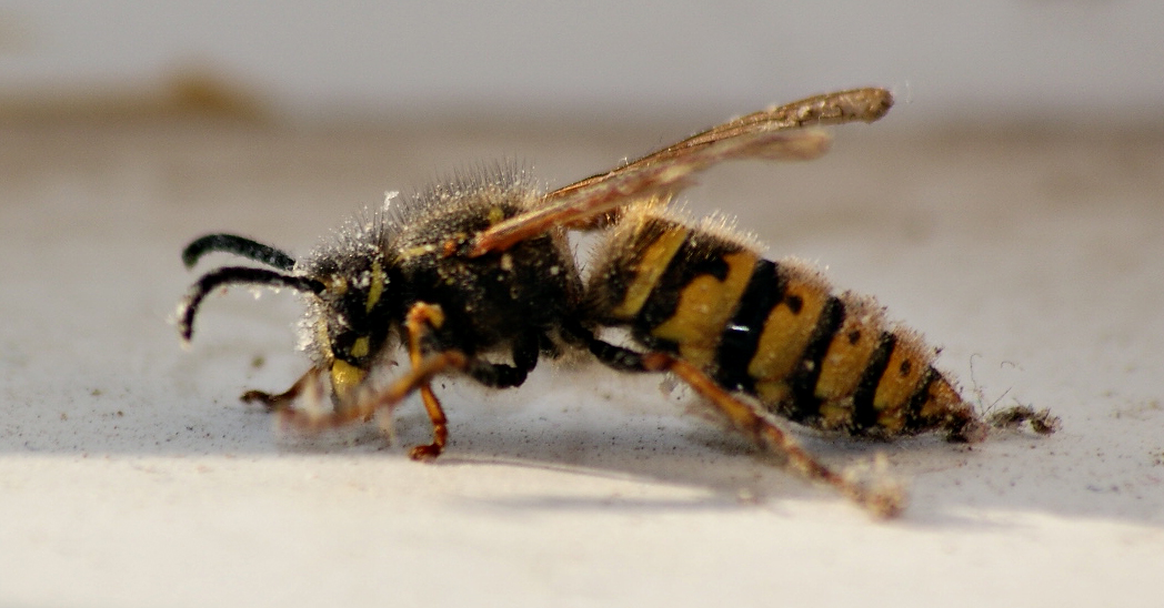
Clinical manifestation: bee colonies are rapidly deteriorating - bees are crawling with spread wings; they cannot fly; are falling on the ground with mildly shriveled abdomen. Infected bees survive one day at most.
Treatment: no treatment or therapy. Bee colonies are to be destroyed on the spot and honeycomb molded into wax. |
New additions
Now you can have all the bee treatment details with you,with bee diseases app from google play. |
Diagnosis: by isolating pathogens from hemolymph on a nutrient medium (agar, broth), coloring (aniline colors) and then by microscopic examination of an isolated pathogen. Also, by biochemical examinations (liquefaction of gelatin, milk coagulation etc.). The material for laboratory examinations are live bees.
Pathoanatomical changes: bee carcasses are fragile, hemolymph of affected bees has milky-white color.
Epizootology: bees become infected via their tracheal system. Conditions for of disease include swarming and building honeycomb. It affects drones and worker bees. Usually strikes in May and wears down in October.
Etiology: disease is caused by 'Bacillus apisepticus', gram negative bacteria, with oval or ellipsoid shape. Anaerobe, doesn't create spores, is affected by temperature. Will die off in the sun in 7h, in 73c in 30min and at 100c in 3min. Will stay active in a bee carcass for 1 month.
Parathypus
Clinical manifestation: bees become weak, and cannot fly. They get liquid, light-brown feces (can be spotted at the bottom of the hive) with swollen abdomen. The disease itself can last from one to a few days (8-15). The bee mortality rate can vary depending on weather conditions - the weather conditions can lower or even sometimes remove the disease, especially if hygiene measures are implemented.
Treatment: if there is no natural recovery (due to weather and good pasture) the following steps are taken: sugar syrup treatment, antibiotic therapy (based on antibiogram), disinfection of all equipment, removing frames with honeycombs stained with feces or moving bees to clean hives. Prevention: destroying insects which can transfer microorganisms to bees, replacing the low quality honey with sugar syrup for better nutrition.
Diagnosis: by isolating pathogens from the hemolymph. Antibiogram is also required. Differential diagnosis: nosemosis (excluded by microscopic examination of intestinal contents (absence of spores of Nosema apis)) and dysentery (examination of intestinal contents (absence of bacteria and undigested pollen))
Pathoanatomical changes: swollen abdomen, liquid intestinal contents, pathohystologycal changes on intestinum.
Epizootology: bees become infected by ingestion of infected food and water. It's caused most often by Bacterium paratyphi alvei. Other conditions are also important: weather conditions during winter, lack of water, weak population. The spread of contagion is done via drones and other bees from different hives, beekeepers and honeycomb frames. Usually occurs in February and march, but period from April to September is not excluded either.
Etiology: caused by 'Bacterium patatyphi alvei' - a round shaped, short gram negative stick, does not create spores, mobile, breeds well on usual nutrient mediums (agar, broth). Biochemical features: fermenting with gas creation, does not dissolve milk, weak to create indol. Cannot artificially infect rats, mice, rabbits and guinea pigs.
Bee dysentery
Clinical manifestation: a lot of liquid excrement can be found in all places in hive (frames, walls, floor). It has scent of floor or cat feces. Bees can't fly, they crawl around the floor (often stained with excrement). They become sensitive to cold and die. The bees themselves are aroused and are crawling everywhere in hive. Prognosis is good if the disease is detected in early stages.
Treatment: no medications are used in therapy of bee dysentery, instead factors that caused the disease are dealt with: bad food should be replaced with sugar syrup, hive should be warmed by placing isolation or a heat source. If there are bee carcasses they should be burned, frames with honey comb are to be cleaned, and washed with hot water. The same should be done with the hive. Honey from the nest should be boiled. If the bee community is heavily affected it should be destroyed, if not, the queen bee should be replaced and the sick bees should be removed. Prevention: the hive should not wait the winter with an old queen bee, or a weak community itself. The hive should not be moved during winter; leave enough food for the winter (at least 7.5kg), check on hive regularly.
Diagnosis: along with confirming clinical symptoms, microscopic examination should be done to confirm absence of 'Nosema apis' from intestinum.
Pathoanatomical changes: bee abdomen is swollen and full of excrement (can be easily ejected if abdomen is pressed), hitinose membrane becomes more transparent
Epizootology and etiology: disease occurs during winter, it's not caused by any specific microorganisms. but by a group of unspecific factors: climatic (long winters, low temperatures, rapid temperature changes), absence of cleansing flights, overeating (anxiety (often caused by noise or pests)), low quality nutrition, lack of water or effect of toxins. All these factors lead to retention of food in bee's intestines of which causes problems in digestion and dysentery. Residual food can also lead to the appearance of some pathogenic microorganisms.
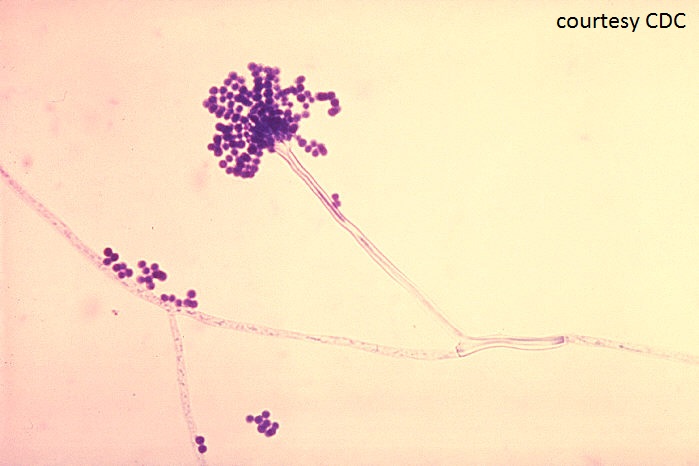
Aspergillosis
Clinical manifestation: disease manifests itself on bees 2 weeks after it appears on the brood. It should be noted that the disease is far easier to spot on the brood than on the bees. Bees are aroused, they become numb and can't fly. They are trying to take off, but they fall on the ground and continue to rapidly move their legs, mouth apparatus and abdominal extremities only to die off after a couple hours.
Treatment: this disease is a zoonosis (it can affect humans) and thus is not treated. If this disease is discovered a veterinary inspection should be contacted immediately in order for hives to be safely destroyed (most often by burning). Bee keepers should not be worried though, most states are refunding damage caused by this disease. When bees are destroyed, hives can be cleaned mechanically and with disinfectants. After that they are washed with hot water and are left to dry. Honey from supper can be used in the confectionery industry after boiling (and mixing with water). During all these times protective equipment should be used (mask, suit and gloves). Note-regardless of what I just wrote here (these are the opinions of the scientific community) I would proceed to burn it all nonetheless since this disease is a zoonosis. Always bear in mind the fact that if any mistake is made you too can get infected.
Diagnosis: microscopic or via nutritive medium. For microscopic examination parts of bee (or larvae) carcasses are taken and sunk into 50% solution of glycerin or NaCl. After that, the sample is observed natively (directly under the microscope). For examination with the nutritive medium special mediums are used (Sabouraud agar). Again when working with A. flavus, care should be taken since it causes complicated pneumonia in humans.
Pathoanatomical changes: spores are germinating from intestinum and destroying the local tissue . Muscles are white and soft.
Epizootology: Favorable factors for disease are high moisture in hive and weak societies. Spores are found everywhere in the hive and on the bees (they can thus spread disease to another hive). Bees are infected by ingesting spores.
Etiology: Fungus Asperegilus flavus Link causes disease. Its conidiospores are colorless with 400-1000 x 5-15 micrometers with their sterigmas. Spores can separate themselves from sterigmas, they are green or yellow in color and are from 2x3 to 5x7 micrometers in size. They can last 30min on a temperature of 60c. Disinfection: 1% sublimat, 5% fenol, 5% formaline.
Paralysis
Acute:
Clinical manifestation: bees are dying almost instantly with symptoms similar to that of chronic manifestation (see below).
Treatment: Same as chronic manifestation. There is no specific therapy, only symptomatic. Bees are administered sugar syrup with the addition of vitamin C, nicotine acid, proteins and saccharides. Substitution of the queen bee is recommended along with induction of bees resistant to the virus. Some experts also recommend spraying bees themselves with sugar syrup in order to stop the virus.
Diagnosis: Based on laboratory examinations: histology exam of bee's intestinum and brain (citoplasmatic basofilic inclusions), artificial infection of healthy bees with material derived from sick bees, test with (resistant) rabbit serum, with electronic microscope and isolation on nutritive medium.
Pathoanatomical changes: similar to chronic manifestation (below).
Epizootology: Latent infection. The virus is excreted by salivary glands, is transmitted among bees by pollen.
Etiology: Caused by a virus from "Picorna" family. Similar to the virus of Baggy brood, he is different from it with his antigen structure. 30nm long, hexagonal shape. Stable when the pH is less than 4.
Chronic:
Clinical manifestation: bees are extracting smaller black bees from a hive, that are resisting and are trying to get back into the hive. It can be wrongly deduced that those are strayed bees from other hives. The bees who are extracting the black bees also start shaking and in few days change color and lose the ability to fly. Other types of symptoms
include shaking with spread wings, legs and antennas. They can’t fly, the abdomen is swollen due to expanded honey bladder which is filled with liquid containing virus. Sick bees are usually gathered in upper parts of the hive above honey combs. They are unable to take off and even if they do they fall in front of the hive gathering in groups, sometimes in hundreds trying to take off. Due to lack of nutrients they starve. Whatever the clinical signs, bees die in 5-7 days. Occurs in winter or at the beginning of the spring.
Treatment: Same as acute manifestation in bees.
Diagnosis: same as acute.
Pathoanatomical changes: swollen black abdomen without hair, sometimes covered with sticky goo with bad odor (rotten fish). White liquid mass can be found in the rectum. With histology examination of brain cells citoplasmatic basofilic inclusions can be found in brain.
Epizootology: It is assumed that the virus is transmitted during "social feeding" of the bees. Disease occurs in summer and stops before fall. Within the apiary usually just a few communities get ill, from year to year (that proves that some bees have genetic affinity for the disease.)
etiology: Disease is caused by RNA virus of ellipsoid shape (30-65nm lenght, 22nm width). Virus is resistant to chloroforme and ether, but is sensitive to higher temperatures. It can survive on 4 degrees Celsius for 4 weeks. It is assumed that that the longer particles are more virulent because they carry more complete genetic information.
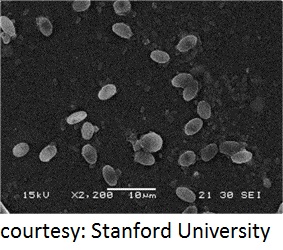
Nosemosis
Clinical manifestation: acute form-high numbers of dead bees at spring. The abdomen is swollen because of residual undigested food. Feces is yellow-grayish and liquid (diarrhea). Bees are unable to take off, are aroused, wandering in circles, gathering in one place and dying. Chronic form - at fall with faint symptoms (or no symptoms at all). Lasts long.
Treatment: On dry and sunny terrain- fumagilin antibiotic. Prophylactic measures-cleaning and disinfection of the honeycomb Microscopic examination of hives in spring and at the fall-regardless of bees' health condition. If detected, this disease is to be reported by law in most countries
Diagnosis: live or dead bees are sent for examination (30-100) properly packed with
corresponding documentation. Microscopic exam: with microscopic sample - bee abdomens are placed in test tubes with saline solution and mixed with glass stick. We then use the glass stick to place the sample on a microscopic plate. We then use a big magnification or x100 (with cedar oil). Spores are of ellipsoid shape like grains of rice.
Pathoanatomical changes: swollen abdomen with undigested food. Pathological changes in epithelium cells on malpighi vessels.
Epizootology with ethiology: The parasite attacks worker bees, drones and the queen bee. It's situated at mid-gut in epithelium cells. Bees are infected by spores (30.000-50.000 are located in intestinum). They can survive 1 year in feces, 2-4 months in honey, a few weeks in free environment, 15-32h at sunlight they are most often found in water, food and feces. They are transferred by several means: moving frame with infected feces, merging diseased with healthy population, with apicultural tools, when bees fly from hive to hive, by ingestion of infected honey.
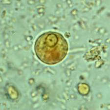
Amebosis
Clinical manifestation: bees die mostly without any clinical symptoms - first older then younger bees. Community weakens - honey production drops. Stronger symptoms would include diarrhea with strong odor, easily comes out - other bees are sprayed too. Despite this, bees can still fly. Weaker symptoms: constricted abdomen, crawling with trembling wings. Occurs in spring.
Treatment: removal of older bees from swarm (disease carriers), artificial swarming 2 weeks before main pasture. Prophylactic measures - examination of society before winter. Most medications haven't shown satisfactory effects.
Diagnosis: samples are taken from live infected bees and placed on microscopic plate. The sample is then colored with gentian violet or methylen blue color (can be seen as a round shell). In summer bees vegetative forms can be detected - we place the malpighi vessels on a microscopic plate and push out their contents (with other smaller plate-also add a drop of water). We can then easily detect vegetative forms.
Pathoanatomical changes: malpighi vessels have a shiny look with necrotic dots.
Epizootology and etiology: caused by amoeba 'mallpighella mellifice prell'. Its development cycle is entirely inside the bee and lasts 24-48 days. It has two forms vegetative and cysts.
Acarosis
Clinical manifestation: bees are unable to take off, shaking, jumping (sometimes with damaged wings); they die while exhaling (in expirium). Generally: number of bees is lowered, dead bees in and around hive.
Treatment: smoke compound with phenothiazine. Smoking 2x a year (spring/fall) - 3x in 10 days at dawn or dusk. Note-in some countries bees with acarosis are not treated but extinguished (by law). The state is refunding damages. Consult a local veterinarian for details.
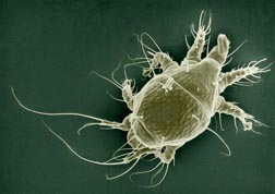
Diagnosis: bees abdomens are placed in test tubes with saline solution; mixed and examined under microscope in search for this parasite: light-yellow color with oval shape and with 4 pairs of legs covered with little hair.
Pathoanatomical changes: damaged wings, the bees die while exhaling - in expirium.
Epizootology and etiology: Caused by 'Acarapis woodi'. This parasite attacks worker bees, drones and the queen bee. Feeds on hemolymph. It invades the bee's respiratory system through the first pair of stigmatic openings. It develops in the first pair of tracheas, rarely on the root of the wings. The fertilized female lays 6-7 eggs. Development cycle 2-3 weeks. It parasites on live bees. When the bee dies it comes off and attaches itself on another bee. Infection among live bees occurs when they come in contact with each other via thoraxes.
Treatment: smoke compound with phenothiazine. Smoking 2x a year (spring/fall) - 3x in 10 days at dawn or dusk. Note-in some countries bees with acarosis are not treated but extinguished (by law). The state is refunding damages. Consult a local veterinarian for details.
Diagnosis: bees abdomens are placed in test tubes with saline solution; mixed and examined under microscope in search for this parasite: light-yellow color with oval shape and with 4 pairs of legs covered with little hair.
Pathoanatomical changes: damaged wings, the bees die while exhaling - in expirium.
Epizootology and etiology: Caused by 'Acarapis woodi'. This parasite attacks worker bees, drones and the queen bee. Feeds on hemolymph. It invades the bee's respiratory system through the first pair of stigmatic openings. It develops in the first pair of tracheas, rarely on the root of the wings. The fertilized female lays 6-7 eggs. Development cycle 2-3 weeks. It parasites on live bees. When the bee dies it comes off and attaches itself on another bee. Infection among live bees occurs when they come in contact with each other via thoraxes.
Bee poisoning
Lower number of worker bees, honey contamination.
Poisoning with chemical agents: most often insecticides and smoke from industrial complex.
Clinical manifestation: worker bees die far out of a hive or in front it. They have spasms, are staggering and falling with spread wings.
H.c.h. poisoning: bees "clean" themselves, open their mouth, fall on the back or on the side. They have a constricted abdomen. Community weakens.
Treatment: destroying dead bees, dead brood, feeding with sugar syrup.
Diagnosis: in toxicology lab- 100g of dead bees with frame and anamnestic data. It's best to keep material at 40 degrees Celsius (that should be delivered within 24h).
Pathoanatomical changes: in intestinum - grains and ulcerations.
Poisoning at pasture
Poisoning with pollen: buttercup, horse chestnut, bear onion: at complexes with those plants or when other plants are late-blooming, so bees use the bad ones instead.
Buttercup: aroused, cant take off, on back, constriction.
Horse chestnut: weakness, sometimes black with swollen abdomen.
Poisoning with "lime pasture": paresis, paralysis, fall far from hive.
Treatment: moving apiaries some place else, reaping of buttercup, not letting them on pasture.
